Volume 6.1 (Spring 2022)

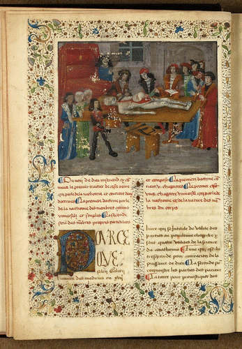
Anatomical Icon: Dissection Scenes in Manuscript and Print, circa 1350–1550 Taylor McCall
By the time Andreas Vesalius (1514–64) created his energetic depiction of a woman’s dissection to grace the front of his anatomical masterwork On the Fabric of the Human Body (1543), the iconography of the dissection scene—a corpse laid on a table surrounded by a crowd of gesticulating men, one wielding a knife, overseen by a lecturer reading from a text—had become firmly emblematic of the field of anatomy. The image appealed at once to educated, professional medical practitioners as well as to members of the general public, increasingly hungry for accessible medical texts, signifying human dissection’s arrival as a central component in the study of medicine. While this may seem natural to modern viewers, human dissection was not performed at all until the late Middle Ages and it took two centuries to become an acceptable part of medical curricula. By delving into the origins of the iconography of the dissection scene in manuscript and print, this essay will tell the story of human dissection in medieval Europe, from the first demonstration in Bologna in the early fourteenth century, to the slow spread of the practice throughout European universities, and arriving at its eventual establishment as an integral part of the practice of medicine by Vesalius’s time. Dissection scene images, which began as a luxurious addition to manuscripts seen only by a small amount of wealthy and educated viewers, became one of the most popular images ever printed as Vesalius’s book soared to fame, reflecting the growing acceptance of the practice.
An Allegory of Wax Anatomies: André-Pierre Pinson Charles Kang
This essay examines the ambiguous position of anatomical models on the spectrum between art and science in late eighteenth-century France. Central to my inquiry is André-Pierre Pinson (1746–1828), originally trained as a surgeon but active primarily as a specialist of polychrome anatomical models in wax. I show how he tried to negotiate his professional identity in settings that were more closely associated with an artistic career in late eighteenth-century Paris: he engaged with the Royal Academy of Painting and Sculpture (Académie royale de peinture et de sculpture), participated in the independent exhibition known as the Salon de la Correspondance, and eventually produced works for the private collection of Louis Philippe Joseph d’Orléans (1747–93). Pinson’s involvement in these settings suggests a desire to have his model-making expertise aligned with artistic production, or perhaps even to venture into conventional art making. To demonstrate Pinson’s sophisticated awareness of his boundary crossing, I focus on the Circulatory System of a Newborn, produced for Orléans in 1789. It thematizes an important contemporary technique of producing wax anatomies, so as to address it conspicuously as a proper subject matter of the work. At the same time, the work aspires to an ideal image, by presenting an imaginary result that the technique would have achieved if it had been devoid of imperfections. This play on the practice of anatomical model making and its limitations makes the Circulatory System an allegory, an artistic device of conveying a complex concept by means of a figural representation.

Anatomy at Large: Caspar Wistar’s Models M. M. A. Hendriksen
By the late eighteenth century, anatomical models were a relatively common phenomenon in European universities, medical colleges, and private collections. Usually made from wax or plaster and often approximately life-sized, they functioned as both educational tools and prestigious objects. Yet in the young American city of Philadelphia, professor of anatomy Caspar Wistar (1761–1818) decided he needed something different for his quickly expanding classes than the models he had seen while studying in Europe. He collaborated with the sculptor William Rush (1756–1833) and collector and artist Charles Willson Peale (1741–1827) and his son, the artist Raphaelle Peale (1774–1825). Together they created larger-than-life models of parts of the human head and neck, using an innovative mix of materials such as papier-mâché, wood, wax, and metal. The models were so well made that they were used for teaching into the twentieth century. This essay starts with a visual and material analysis of one such model and subsequently places it within the context of objects, people, practices, and discourse surrounding it to cast light on the importance of artisanal knowledge and skills for the development of Philadelphia as a center of medical education in the early nineteenth century.
A Cyclops, a Stone, and a Four-Breasted Woman: Drawing Anatomical Knowledge Katherine M. Reinhart
Drawings have a long history communicating new ideas, and in the early modern period, they were mobilized by a new form of institution—the scientific society—which emerged as a permanent structure to facilitate novel modes of pursuing and producing anatomical knowledge. This collaborative approach engendered new techniques for communicating and circulating knowledge among investigators, including innovative visual and material practices. Institutions such as the Royal Society of London and the Académie royale des sciences in Paris produced hundreds of drawings, by both artists and philosophers, in the course of their pursuits to understand the functions, structures, and diseases of the body. From depictions of bladder stones to monstrous births, drawings not only recorded anatomical investigations but often stood in for anatomical objects themselves. This essay explores how these early scientific societies created and used drawings as an important tool in their work, particularly in the study of human anatomy. From drawn accounts of dissected cadavers to recording anatomical anomalies, drawings circulated in society meetings, were debated by members, and were recorded in the institution’s minutes. This essay focuses on the materiality of drawings and their ability to preserve and perpetuate anatomical knowledge in ways that ephemeral specimens were not able to match. Based on unstudied archival drawings, this essay contends that image-making was a central component to the study of human anatomy in early scientific societies and that these drawings were a crucial tool in the process of anatomical discovery and knowledge production during their early years.

Mistress Mummy: Dissecting the Flesh-and-Blood Venus Margaret Carlyle
The Venus or female nude has been a long-standing motif in art history, from the ancient Greek marble statue of Venus de Milo to Sandro Botticelli’s fifteenth-century painting of her “birth.” By the eighteenth century, a new kind of Venus emerged in Europe’s museums that sat at the crossroads of art and anatomy: a lifelike woman made of colored wax. Donning a pearl necklace, long hair, and a touch of rouge, she was invariably displayed lying on satin sheets inside a glass display case for the titillation of a curious public. Behind her pleasing aesthetic, however, was an anatomy lesson: her life-size inner organs could be removed and replaced at will, in an almost divine act that mimicked the art of human dissection.
While the “Anatomical Venus” was an important figure who dazzled audiences in such enlightened cities as Vienna, London, and Florence, we know much less about her bloodier, messier counterpart who lay on the dissecting table: the female corpse. This essay makes a case for the importance of the female corpse—what I will call the “flesh-and-blood Venus”—to anatomical inquiry. By looking at dissections of such Venuses behind closed doors, before auditoriums of eager students, and in medical imagery, this essay shows that the flesh-and-blood Venus functioned less as a spectacle than as a testament to the anatomist’s disciplinary mastery. Examples of a mummified mistress, a teenager found lifeless in a ravine, and a woman who died in the ninth month of her pregnancy will show how the female corpse was central to new forms of anatomical inquiry in such fields as childbirth and forensics. The flesh-and-blood Venus provided anatomists with a unique research site to advance new knowledge claims while displaying their disciplinary expertise.
A Hairy Tale: Eighteenth-Century Strands of Albinism and Race Sarah-Maria Schober
Besides his extraordinarily well-known collection of human skulls, the German anatomist and anthropologist Johann Friedrich Blumenbach (1752–1840) was an avid collector of human hair. Largely overlooked by historical research so far, Blumenbach’s samples of hair prove highly significant in the formation of eighteenth-century ideas about race. Among the samples, seven specimens of albino hair stand out. Up until the late eighteenth century, albinism had been understood as a singularly extra-European phenomenon of “white Negroes.” As such, albinism became deeply embedded in the evolution of theories of race. The evidence collected by Blumenbach through hair samples pointed, however, to another explanation: for Blumenbach, albinism was an illness and did not constitute a different human variety. Therefore, it did not challenge his theory about five human races.
This article will argue that Blumenbach’s supposedly more “scientific” way of dealing with people with extraordinarily fair skin and hair did indeed foster his system of racial thought. By splitting up his observations on anatomical things like hair and skulls into the two realms of anthropology and anatomy, Blumenbach succeeded in bypassing the “problem” of the “white Negro” by separating it as an illness apart from his attempt to categorize humans anthropologically. The story of Blumenbach’s albino hair samples therefore fits perfectly into the context of the formation of the life sciences in the eighteenth century. It provides proof not only of a disciplinary differentiation between anatomists and anthropologists but also of the ongoing racial entanglement of the sciences.